


1
/
of
5
ECG 7-Step Plan – Interpretation Reference Card
ECG 7-Step Plan – Interpretation Reference Card
2 reviews
Regular price
£3.50 GBP
Regular price
Sale price
£3.50 GBP
Unit price
/
per
Tax included.
Couldn't load pickup availability
SKU:CARD-7STEP
- Two-sided ECG interpretation reference card following a simple 7-step structured approach. Includes:
- Heart rate
- Rhythm
- Axis
- P waves and PR interval
- QRS complex and QT interval
- ST segment changes
- T wave abnormalities
- Front side covers the full 7 Step Plan, including:
- Assessment of heart rate with normal ranges
- Normal 60–100 bpm
- Tachycardia >100 bpm
- Bradycardia <60 bpm
- Rhythm analysis including regular, irregular, regularly irregular, and irregularly irregular rhythms
- Axis interpretation including normal, left, right, or extreme axis
- Assessment of P waves and PR interval including:
- Presence of P waves
- Relationship of P waves to QRS
- QRS following each P wave
- P wave morphology
- Normal PR interval 120–200 ms
- Assessment of QRS complex and QT interval including:
- Normal QRS 80–120 ms
- QRS morphology
- Normal QTc 350–440 ms (males) and 460 ms (females)
- ST segment evaluation including:
- Isoelectric baseline
- Recognition of elevation or depression and possible ischaemia
- T wave assessment including:
- Normal shape
- Inversion
- Increased amplitude
- Assessment of heart rate with normal ranges
- Reverse side includes common ECG abnormalities, including:
- Absolute bradycardia <40 bpm
- Prolonged PR interval >200 ms
- Short PR interval <120 ms
- Narrow QRS <80 ms
- Broad QRS >120 ms
- Prolonged QTc >440 ms (males), >460 ms (females), >500 ms (high risk)
- Reverse side also provides diagnostic clues for each abnormality, including associations with:
- Sick sinus syndrome
- Heart block
- Electrolyte imbalances
- Drug effects such as beta-blockers, calcium channel blockers, digoxin
- sodium channel blockers
- Bundle branch blocks
- Ventricular rhythms including VT and PVCs
- Pre-excitation (WPW, LGL)
- Hypokalaemia, hyperkalaemia, LQTS
- Myocardial infarction
- T wave patterns and associated causes including:
- Peaked
- Inverted
- Biphasic
- Hyperacute
- Flattened
- Camel hump patterns
- Designed as a study and revision aid for students and clinicians learning structured ECG interpretation.
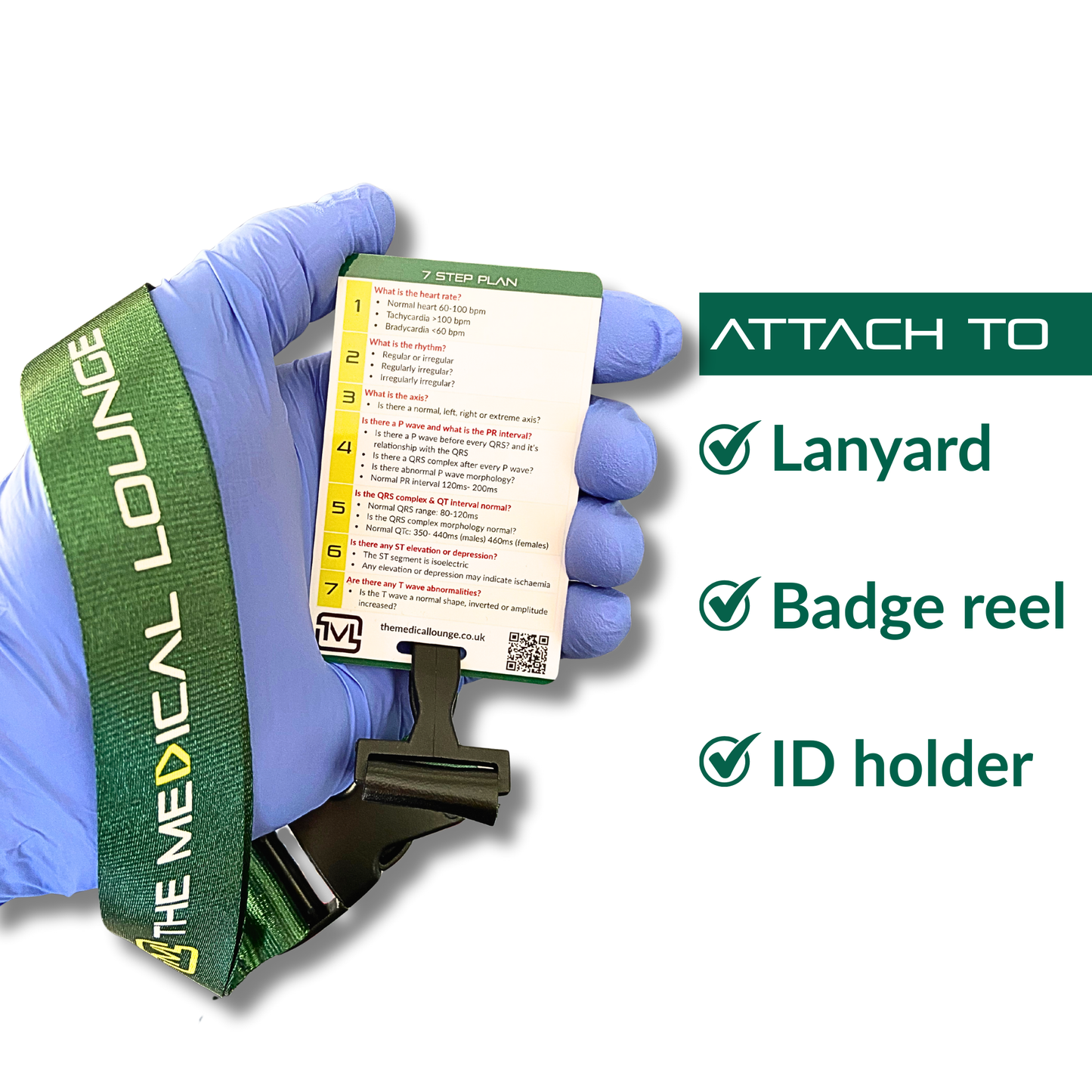
- Pocket-sized 86 × 54 mm laminated card with a durable elongated slot for lanyards, badge reels, or ID holders.
- High-quality print, fully encapsulated in transparent laminate for everyday use.
For educational and reference use only.
Share


R
R.W. Great cards for a lanyard
A
Anonymous Very good course