Abdominal pain is one of those complaints you’ll see all the time on the ambulance. It ranges from “bit of a dodgy kebab” to “this person needs a surgeon yesterday” and at 3am, they can all look suspiciously similar.
A structured abdominal assessment helps you sort the benign from the time-critical. This blog walks through a simple I-A-P-P approach (Inspect, Auscultate, Palpate, Percuss) and shows how it links directly to our Abdominal Assessment pocket card, so you’ve got the key prompts in your hand, not just in your memory.
Before you start
Before you get hands-on:
-
Ensure exposure with dignity – lift clothing appropriately, use blankets, close doors/curtains.
-
Position the patient lying flat, knees slightly bent if possible (reduces muscle tension).
-
Ask about pain location and severity first – you don’t want to prod the worst bit without warning.
-
Warm hands, explain what you’re doing, and keep an eye on their face for discomfort.
Once you’re set, follow the sequence on the front of the card: I – A – P – P.
I – Inspect
You start with inspection because it’s non-invasive and already gives you a lot of information.
On the card, the Inspect section reminds you to look for:
-
Scars – previous surgery (laparotomy, C-section, hernia repair) can hint at adhesions, bowel obstruction, or past pathology.
-
Bruising –
-
Grey-Turner sign (flanks)
-
Cullen’s sign (around the umbilicus)
Both can be associated with retroperitoneal bleeding or severe pancreatitis.
-
-
Distension – the classic “6 F’s”:
Fat, Fluid, Flatus, Fetus, Faeces, Fibroids.
You’re trying to decide if this abdomen is simply “generous” or acutely distended and tense. -
Stomas – note site, type, output. A bag full of blood or no output in someone with pain is significant.
-
Visible masses – look for site, size, shape, and whether it appears pulsatile.
Visually, ask yourself: Does this abdomen look soft and relaxed… or tight, shiny, bruised, and worrying?
A – Auscultate
Auscultation comes before palpation so you don’t alter bowel activity.
The card prompts you to assess:
-
Bowel sounds –
-
Present / normal
-
Absent
-
Tinkling (classically high-pitched, can suggest obstruction)
-
Hyperactive (often early obstruction or irritation)
-
-
Bruits – over the aorta, renal, and iliac areas. A whooshing sound can suggest turbulent flow (e.g. aneurysm or stenosis).
-
Friction rubs – over the liver or spleen (rare, but if you hear one, it’s a big clinical clue to inflammation of those organs).
In pre-hospital practice, you won’t always have a quiet environment for detailed auscultation, but when you can, it adds useful pieces to the puzzle.
P – Palpate
Now you move to palpation, starting gently and away from the area of worst pain.
The card breaks palpation into three key ideas:
-
Light and deep palpation
-
Start superficially to identify tenderness and to gauge the patient’s pain response.
-
Progress to deeper palpation if tolerated to feel for masses or organ edges.
-
-
Guarding vs rigidity
-
Voluntary guarding – the patient tenses because it hurts or they’re anxious. It often eases if you distract or reassure them.
-
Involuntary rigidity – the muscles are board-like, regardless of the patient’s effort. This suggests peritonitisand is a big red flag.
-
-
Masses and organomegaly
-
Is that lump mobile or fixed, tender or non-tender?
-
Can you feel an enlarged liver or spleen?
-
Ballot the kidneys (press from the back and feel anteriorly) if flank pain is part of the picture.
-
Watch their face the whole time: sometimes the patient will grimace before they say anything.
P – Percuss
Percussion can feel a bit “old school”, but it’s an efficient way to get more information quickly.
The card focuses on three percussion findings:
-
Tympany – a drum-like sound, usually from gas-filled bowel.
-
Dullness – suggests fluid, solid organs, or masses.
-
Shifting dullness – percuss from the midline outwards, then repeat with the patient turned slightly on their side. A change in dullness can indicate ascites.
In the back of your mind, you’re asking: Is this mostly gas? Mostly fluid? Or something solid that shouldn’t be there?
Red flags you cannot ignore
At the bottom of the card is a Red Flags section, essentially the “do not down-triage this” list.
Key red flags include:
-
Guarding or rigidity (especially involuntary)
-
Rebound tenderness
-
Distension with signs of shock (tachycardia, hypotension, cool peripheries)
-
Pulsatile abdominal mass
-
Haematemesis or melaena
Any of these should prompt you to think time-critical pathology: perforation, peritonitis, bowel obstruction, ruptured AAA, significant GI bleed. In the pre-hospital world, that usually means:
-
Early alert / pre-alert
-
Rapid transport to an appropriate destination
-
Good ATMIST or equivalent, including onset, red flags, and haemodynamics
Associated symptoms: don’t forget the rest of the history
Abdominal pain rarely comes alone. The card gives a quick reminder to explore:
-
Nausea and vomiting
-
Change in bowels – diarrhoea, constipation, blood or mucus
-
Jaundice
-
Urinary symptoms – dysuria, frequency, retention
-
PV/PR bleeding
This is where you weave in SOCRATES, medication history, alcohol use, anticoagulants, previous episodes, and red flag systemic symptoms (fever, weight loss, rigors).
Clinical pearls: small details that change your thinking
The Pearls / Clinical Notes section on the card distils some high-yield principles that are easy to forget under pressure:
-
Pain before vomiting → surgical cause more likely
(Think obstruction, appendicitis, perforation.) -
Pain migrating to the right iliac fossa → appendicitis
Classically starts peri-umbilical and then localises. -
Sudden, severe onset pain → consider AAA or perforation
Especially in older patients or those with vascular risk factors. -
Always consider pregnancy
Any patient of child-bearing potential with abdominal pain = think ectopic pregnancy until proven otherwise.
These aren’t absolute rules, but they nudge you to keep serious pathology on your radar.
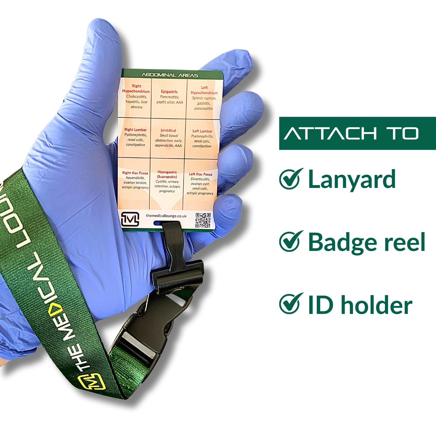
Mapping pain: abdominal areas and likely causes
The reverse side of the card breaks the abdomen into nine areas and lists common pathologies in each region, a handy way to link “where it hurts” to “what might be going on.”
Examples:
-
Right hypochondrium – cholecystitis, hepatitis, liver abscess
-
Epigastric – pancreatitis, peptic ulcer, AAA
-
Left hypochondrium – splenic rupture, gastritis, pancreatitis
-
Right lumbar / left lumbar – pyelonephritis, renal colic, constipation
-
Umbilical – small bowel obstruction, early appendicitis, AAA
-
Right iliac fossa – appendicitis, ovarian torsion, ectopic pregnancy
-
Hypogastric (suprapubic) – cystitis, urinary retention, ectopic pregnancy
-
Left iliac fossa – diverticulitis, ovarian cyst, renal colic, ectopic pregnancy
In practice, you can:
-
Ask the patient to point with one finger to the main site of pain.
-
Match that region to the grid on the card.
-
Combine site + character of pain + associated symptoms + exam findings to build your differential.
It’s not about making a definitive diagnosis curb-side, but about recognising patterns and identifying those who need urgent imaging, surgery or medical review.
Bringing it all together on the truck
For student paramedics, abdominal assessment can initially feel like a lot of separate pieces. The aim of the Abdominal Assessment card is to give you:
-
A clear I-A-P-P structure to follow
-
Prompts for what to look, listen, feel, and percuss for
-
A quick red flag checklist
-
A visual abdominal map linking pain location to likely causes
Use it during placements, OSCE practice, and real jobs until the sequence becomes second nature. Then the card becomes what it was designed to be: a fast, reassuring prompt when you’re tired, it’s 4am, and yet another patient says, “It’s just a bit of tummy ache, love.”